Showing posts with label Brain. Show all posts

Saturday, April 30, 2016

Creating False Memories

Neuroscientists in France have implanted false memories into the brains of sleeping mice. Using electrodes to directly stimulate and record the activity of nerves cells, they made artificial associative memories that lasted while the animals slept and then influenced their behavior when they woke up.
Manipulating memories by messing with brain cells is nothing new in neuroscience labs. In 2014, one team of researchers used a technique called optogenetics to label the cells encoding fearful memories in the mouse brain and switch them on and off, and another used it to identify the cells encoding positive and negative memories, so that they could change positive memories into negative ones and vice versa.
This new work, published March 9, 2015 in the journal Nature Neuroscience, shows for the first time that artificial memories can be put into the brains of sleeping animals. It also gives scientists more details about how populations of nerve cells encode spatial memories and the important role that slep plays in making these memories stronger.
Karim Benchenane of the French National Centre for Scientific Research in Paris and his colleagues put electrodes into the brains of 40 mice, targeting the medial forebrain bundle(MFB), a component of the reward circuitry which has at least 3 different cells types that encode the memories needed for spatial navigation. Then they left the mice to explore their environment and monitored the responses of their neurons to identify place cells, each of which fired when one of the animals was in a specific location in their surroundings. In one experiment, performed on 5 awake mice, they timed the electrical stimulation of the MFB to correspond with the firing of a specific place cell.
This paired stimulation gave the mice a false memory. The mice linked the MFB stimulation with the specific location encoded by the cell, and thus spent 4- to 5-times more time in that location than two control mice who received MFB stimulation that did not correspond with a place cell firing. Place cells are known to 'replay' their activity patterns during sleep, and this is thought to strengthen newly formed memories.
To research further, the researchers repeated their experiments in 5 sleeping mice. Having already identified place cells while the mice explored their environment, the researchers let them fall asleep, and then paired the firing of each selected place cell in each one with the stimulation of the MFB. Later on, these mice, as well, showed a large preference for that specific location. In contrast, the control mice, who received random MFB stimulation that was not paired with the firing of a place cell wandered around with no liking to any particular place.
Even so, other work shows that false memories can be implanted into the human brain without advanced technology. In a recent study, nearly 3/4 of participants reported having in depth false memories of a crime they didn't commit.
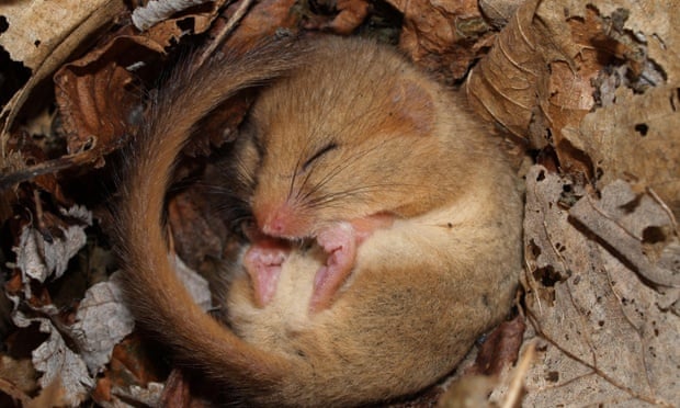
By directly manipulating the brains of sleeping mice, researchers tricked the animals into thinking they had received a reward at a specific place.Photo Credit

The Neurobiology Behind Individuality

When a group of genetically identical mice lived together in the same complex for three months, mice that explored their surroundings more broadly grew more new neurons than those who were less adventurous, according to a study published May 9, 2013 in Science Magazine. This link between inquiring behaviors and the adult growth and development of nervous tissue shows that brain plasticity can be shaped by experience and suggests that this may promote individuality, even in genetically identical organisms.
Scientists have long tired to tackle the question of how individual differences in behavior and personality develop in terms of the way genes and the individual's surroundings interact. "But there is next to nothing [known] about the neurobiological mechanisms underlying individuality" says Gerd Kempermann of the German Center for Neurodegenerative Diseases in Dresden.
A logical way to study this would be to look at how the brain's structure and function change over time (the brain's plasticity). This plasticity is hard to study, though, because it takes place mostly at the synaptic level, so Kempermann and his colleagues decided to look at the growth of new neurons in the adult hippocampus, which can be found easily. Earlier studies have shown that activity increases the adult growth an development of nervous tissue in groups of genetically identical mice, but there were differences between individuals in the amount of neuron growth.
To find out why, Kempermann and his colleagues housed 40 genetically identical female inbred mice in a 5-square meter, 5-level enclosure filled with different sorts of objects made to encourage activity and exploration. The mice were tagged with radio-frequency-infer-red transponders and had 20 antennas in the complex that caught their every movement. After 3 months, the researchers assessed the growth and development of nervous tissue in the mice by counting their fast-multiplying precursor cells, which had been numbered before the study began.
The researchers saw that individual differences in exploratory behavior corresponded with individual differences in the numbers of new neurons generated. "To our knowledge, it's the first example of a direct link between individual behavior and individual brain plasticity," said Kempermann.
However, be cautious about putting all the differences on their surroundings. Even though the mice in the study were genetically the same, they were not behaviorally identical to begin with. It is clear that some variation happens at an extremely early stage, making the mice more or less likely to explore.
These findings could help explain why human identical twins raised in the environment end up with different personalities. Photo Credit

Friday, April 8, 2016

Artificial Skin That can Give People with Prothestics a Sense of Touch

Artificial skin made in a lab can "feel" similar to the way a fingertip senses pressure and could one day let people feel sensation in their prosthesis limbs. Researchers were able to send the touching sensation as an electric pulse to the relative "touch" brain cells in mice.
The stretchy, flexible skin is made of a man made rubber that has been manufactured to micron-scale pyramid like structures that make it extra sensitive to pressure. The researchers sprinkled this pressure sensitive rubber with microscopic cylinders of carbon that conduct electricity very well so that when the material is touched, a series of electric pulses is given off by the sensor. This series of pulses is then sent to the brain cells in a way that mirrors how touch receptors in human skin send sensation to the brain.
To test if the skin could make electric pulses that the brain could respond to, the researchers connected the synthetic skin to a blue LED light. When the skin was touched, it sent electric signals to the LED light, which pulsed in response. The sensors translated that pressure pulse into electric pulses. When the sensors in the skin sent the electric pulses to the LED- very similar to the way touch receptors in real human skin send touch sensation signals to the brain- a blue light flashed.
The researchers added a special protein that cause brain cells to react to blue light and added them to the mice brain cells. The protein let the LED act like a receptor cells in the skin. When the light flashed it sent a signal to the brain that the artificial skin had been touched. This experiment showed that, when the artificial skin was touched, the brain would respond in the same way as brains react to real skin being touched.
The next step would be to the skin in other animals and the eventual goal is to be able to stimulate the human brain and have the skin available for people.

Thursday, April 7, 2016

Is You'r Sense of Taste Connected to You'r Sense of Smell?

Taste is extremely connected with smell. Since our taste buds only detect 5 tastes, smell plays an important part in helping us taste flavors. The 5 categories are: sweet, salty, sour, bitter, and sweet. Now imagine all of the flavors you have ever tasted and subtract any of these five that apply to a certain food. All of that flavor is because you are able to smell! We sometimes take smell for granted, but when we realize how important it is we are able to thank God for what he has given us.

If you want to learn some more click this link

Monday, February 29, 2016

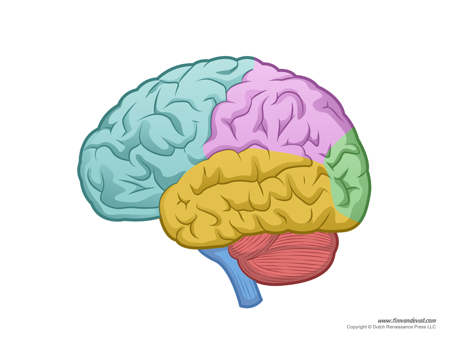
Parts of the Brain (And How They Work)

The brain is a very complex thing. We still don't understand everything about it and how it works, but here's a quick layout:

Light Blue- This area of the brain is called the frontal lobe. It controls the ability to do problem-solving, judgement, and motor function.

Purple- This area of the brain is called the parietal lobe. It controls sensation, handwriting, and body position.

Green- This area of the brain is called the occipital lobe. It controls the brain's visual processing system.

Yellow- This area of the brain is called the temporal lobe. It controls the ability to memorize and hear.

Red- This area of the brain is called the cerebellum. It is responsible for coordination and balance.

Dark Blue- This is called the brain stem. It controls breathing, sleep, and the heart.

There are also other parts of the brain. The cortex is the outer layer of brain cells. It controls thinking and voluntary movements. The basal ganglia are groups of structures in the brain. They arrange messages to send to other parts of the brain.

Photo Credit
Info Credit

Tuesday, May 19, 2015

The science of laughter!

Laughter is usually looked down upon in class but it is actually an interesting thing to study. Did you know that laughter is contagious. When people realized this they started putting "laugh tracks" with their shows which means that you are more likely to laugh at it. For more information on the science of laughter you can click here.